SCIENTIFIC DISCOVERY
With the help of this discovery, we obtained a layer of contracting cardiomyocytes and grew hair in the laboratory (for the first time in the world).
«New data on cellular resources».
We discovered Previously unknown structures (3rd type) involved in skin regeneration processes. For this purpose a special protocol of tissue processing was applied. These structures do not yet have their own common name (the discovery has not yet been evaluated by the international community), and for this reason temporary terms are used.
New data on cellular resources
Structures
- Fig. 1"Capsular structures".
Confocal microscopy. Staining: blue color - Hoechst dye, tropic to DNA; red color - rhodamine phalloidin - dye on cell cytoskeleton; ckit - -green color - stem cell marker. - Fig. 2"Spherical" structures.
Inverted microscope. Rep-50 μm. - Fig. 3"Crystals."
Confocal microscopy. Staining: blue color - Hoechst dye, tropic to DNA; red color - rhodamine phalloidin - dye on cell cytoskeleton; ckit - - green color - stem cell marker. A newborn cell "emerges" from the structure
All kinds of structures were found in various organs and tissues (myocardium, skeletal muscle, cartilage, bone, skin, corneal and conjunctiva epithelium) in different animals (rat, calf, axolotl, lamprey, Danio Rerio fish and human). The mentioned "findings" have a non-random character. The discovery of these structures, to a certain extent, breaks the already established ideas about the ways of tissue proliferation.
"Capsular" and "crystal-like" structures are related to regeneration, and, apparently, are also varieties of previously unknown regional stem cells.
There is more clarity on "spherical structures". Applied (commercial applications of them are being seen now).
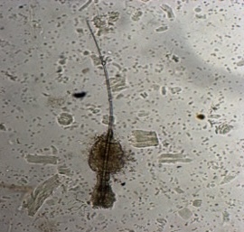
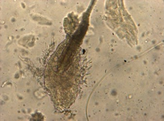
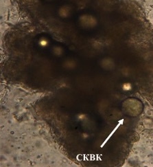
"Spherical structures" have been found in many organs and tissues (skeletal muscle, heart, liver, cartilage, skin) in various animals (rat, rabbit, cow, human, lizards, fish, axolotl, lamprey, etc.). Of greatest interest is skin, in which they are detected mainly in the hair follicle bulb, i.e. where stem cells are located ( Fig. 4.).
Photo to the right: hair isolated from skin by special treatment. Inverted microscopy. "Globular structures" are visible in the bulb.
"Capsular" and "crystal-like" structures are related to regeneration, and, apparently, are also varieties of previously unknown regional stem cells.
There is more clarity on "spherical structures". Applied (commercial applications of them are being seen now).
"Spherical structures" have been found in many organs and tissues (skeletal muscle, heart, liver, cartilage, skin) in various animals (rat, rabbit, cow, human, lizards, fish, axolotl, lamprey, etc.). Of greatest interest is skin, in which they are detected mainly in the hair follicle bulb, i.e. where stem cells are located ( Fig. 4.).
Photo to the right: hair isolated from skin by special treatment. Inverted microscopy. "Globular structures" are visible in the bulb.
All kinds of structures were found in various organs and tissues
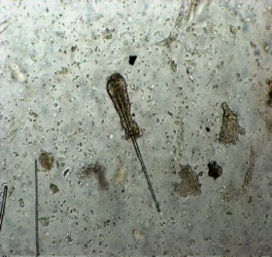
After their isolation and in the process of cultivation, their number and size increase (Fig. 5, a-c). When staining with luminescent dyes (Fig. 5d) on DNA (DAPI dye), it can be seen that almost all the contents of the "ball" consists of nuclear material. In the process of "balloon maturation" a complex reorganization takes place inside them (Fig. e) with the formation of characteristic confocal microscopy showed that in the process of "balloon maturation" (6.a-d) not only the size of structures changes but also their internal "content" - internal structures are formed.
Fig. below (5). A,-d) "Capsule-free" spherical structures at the hair surface, inverted microscope, uv-e.25-40; e) fluorescent microscope, DAPI staining for nuclear material. f)confocal microscopy
Fig. below (5). A,-d) "Capsule-free" spherical structures at the hair surface, inverted microscope, uv-e.25-40; e) fluorescent microscope, DAPI staining for nuclear material. f)confocal microscopy
All kinds of structures were found in various organs and tissues
All kinds of structures were found in various organs and tissues (myocardium, skeletal muscle, cartilage, bone, skin, corneal and conjunctiva epithelium) in different animals (rat, calf, axolotl, lamprey, Danio Rerio fish and human). The mentioned "findings" have a non-random character. The discovery of these structures, to a certain extent, breaks the already established ideas about the ways of tissue proliferation.
"Capsular" and "crystal-like" structures are related to regeneration, and, apparently, are also varieties of previously unknown regional stem cells. There is more clarity on "spherical structures". Applied (commercial applications of them are being seen now).
"Spherical structures" have been found in many organs and tissues (skeletal muscle, heart, liver, cartilage, skin) in various animals (rat, rabbit, cow, human, lizards, fish, axolotl, lamprey, etc.). Of greatest interest is skin, in which they are detected mainly in the hair follicle bulb, i.e. where stem cells are located ( Fig. 4.).
Fig. below (4).: hair isolated from skin by special treatment. Inverted microscopy. "Globular structures" are visible in the bulb.


After their isolation and in the process of cultivation, their number and size increase (Fig. 5, a-c). When staining with luminescent dyes (Fig. 5d) on DNA (DAPI dye), it can be seen that almost all the contents of the "ball" consists of nuclear material. In the process of "balloon maturation" a complex reorganization takes place inside them (Fig. e) with the formation of characteristic confocal microscopy showed that in the process of "balloon maturation" (6.a-d) not only the size of structures changes but also their internal "content" - internal structures are formed.
Fig. below (5). A,-d) "capsule-free" spherical structures at the hair surface, inverted microscope, uv-e.25-40; e) fluorescent microscope, DAPI staining for nuclear material. f)confocal microscopy
Fig. below (5). A,-d) "capsule-free" spherical structures at the hair surface, inverted microscope, uv-e.25-40; e) fluorescent microscope, DAPI staining for nuclear material. f)confocal microscopy
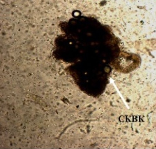
Fig. below (6). confocal microscopy. Staining: blue color - Hoechst dye (DNA dye); red color (rhodamine phalloidin - cell cytoskeleton dye); ckit - green color - stem cell marker. a) staining with "stemness" marker; b) formation of small spherical structures (trazitor cells) inside "spheres".
Fig. 6. confocal microscopy.
Staining:
blue color - Hoechst dye (DNA dye);
red color (rhodamine phalloidin - cell cytoskeleton dye);
ckit - green color - stem cell marker.
a) staining with "stemness" marker;
b) formation of small spherical structures (trazitor cells) inside "spheres".
Staining:
blue color - Hoechst dye (DNA dye);
red color (rhodamine phalloidin - cell cytoskeleton dye);
ckit - green color - stem cell marker.
a) staining with "stemness" marker;
b) formation of small spherical structures (trazitor cells) inside "spheres".
Over time, the number of "balls" in the culture further increases and they are located around the hair in the form of "bunches of grapes" and individual cells emerge from them.At the end of "maturation" small cells* can emerge from the Ball Structures (Fig.7.). The process of "exit" of cells from the spheres ("labor") takes little time, it occurs quite spontaneously and it is difficult to "catch" and fix (Fig.8,9).
Photo series below (7): clusters of spheres near the hair and appearance of small bulges on their surface. inverted microscopy, magnification 40.
Photo series below (7): clusters of spheres near the hair and appearance of small bulges on their surface. inverted microscopy, magnification 40.
Over time, the number of "balls" in the culture further increases and they are located around the hair in the form of "bunches of grapes" and individual cells emerge from them.At the end of "maturation" small cells* can emerge from the Ball Structures (Fig.7.). The process of "exit" of cells from the spheres ("labor") takes little time, it occurs quite spontaneously and it is difficult to "catch" and fix ( Fig.8,9).
Photo series below (7): clusters of spheres near the hair and appearance of small bulges on their surface. inverted microscopy, magnification 40.
Photo series below (7): clusters of spheres near the hair and appearance of small bulges on their surface. inverted microscopy, magnification 40.
Fig.below (8). exit of cells from a spherical structure ("labor"). Inverted microscope.
Fig.below (8). exit of cells from a spherical structure ("labor"). Inverted microscope.
Photo series below (9): cell isolation from the ball of a DanioRerio fish. Confocal microscopy.
Photo series below (9): cell isolation from the ball of a DanioRerio fish. Confocal microscopy.
The number of Globular structures around different parts of the hair increased with different variants of long-term cultivation (in growth medium and in collagen gel).
Photo series below (10): appearance of a large number of "capsule-free" spherical structures inside (a) and around the rod part of the hair (b,c).
Photo series below (10): appearance of a large number of "capsule-free" spherical structures inside (a) and around the rod part of the hair (b,c).
Fig. below (11). different-sized "spherical structures" in collagen gel from which cells are isolated.
Fig. below (11). different-sized "spherical structures" in collagen gel from which cells are isolated.
There are processing modes when "spherical structures" appear inside the hair (the hair shaft), resulting in the formation of "branching hair"! During long-term cultivation, the phenomenon of "budding" and the subsequent development of a "branch" in the hair shaft was discovered. The same hair shafts were involved in this process (Fig.12,13).
Photo series below (12). initiation of "budding" from the rod part of the hair with the appearance of the inside of the hair.
Photo series below (12). initiation of "budding" from the rod part of the hair with the appearance of the inside of the hair.
Photo series below (13): formation of "outgrowth" (twig) in the hair shaft part on the 26th day of cultivation. A) general view of the hair; B) structure of the outgrowth; C) "pedestal" of the outgrowth with spherical structures
Photo series below (13): formation of "outgrowth" (twig) in the hair shaft part on the 26th day of cultivation. A) general view of the hair; B) structure of the outgrowth; C) "pedestal" of the outgrowth with spherical structures
Cultivation regimes were obtained in which hair follicle rudiments were formed from "spherical structures" (Fig.14,15).
Photo series below (14): formation of the hair follicle rudiment.
Photo series below (14): formation of the hair follicle rudiment.
Cultivation regimes were obtained in which hair follicle rudiments were formed from "spherical structures" (Fig.14,15).
Photo series below (14): formation of the hair follicle rudiment.
Photo series below (14): formation of the hair follicle rudiment.
Photo series below (15): forming hair follicle in collagen gel (next to the hair).
Photo series below (15): forming hair follicle in collagen gel (next to the hair).
RESULTS
Preliminary findings:
There are several sources of hair growth: proliferating cells of the bulb; cells (transient) released from capsule-free globular structures inside the hair shaft. Inside the spherical structure, the formation of future cell components (primarily - its nuclear component) takes place.
A brief conclusion on "spherical" structures.
The previously unknown phenomenon of cell replication inside the spherical supracellular structures has been established, which consists in the fact that when signals of physical, chemical and biological origin are received, the mechanisms of cell replication of different levels of differentiation (stem and transient) are activated inside these spherical structures by means of activation of genetic material included at the stage of embryonic development or introduced by means of entosis, with the formation of multiple cellular structures involved in long-range cellular development. Separate and less studied areas of research are "capsule structures" and "crystal structures", which are also involved in regeneration.
+7 999 539 99 96
Src.company@yandex.ru
Src.company@yandex.ru
Saint- Petersburg, 2024
